Radiation and Computed Tomography (CT Scans)
Ionizing Radiation and X-Rays
Ionizing radiation is a form of energy that can interact with other molecules causing the atoms to lose and electron, to form an ionized molecule or ion. X-Rays are a form of ionizing radiation used in medical exams, such as X-Rays and CT scans, that are able to penetrate the body and by interacting with different tissues can produce internal pictures for Radiologists to interpret.
The discovery of X-Rays is credited to Wilhelm Conrad Röntgen, a German physicist, as he was the first to systematically study them. In 1895, his wife’s left hand became the first image of looking inside the human body. At the time, he really didn’t know what the radiation was and named it “X” which is a term that stuck. For his discovery, he was awarded the Nobel Prize in 1901.
X-Rays and CT Scans
X-Ray Computed Tomography or Computed Assisted Tomography, simply known as CAT or CT, was invented in 1971 by Godfrey N. Hounsfield and Allan M. Cormack, who were jointly awarded the Nobel Prize in Medicine in 1979. Thus, CT scans themselves are multiple X-Rays taken as the rotate around the body and then a computer takes the information to produce 3-dimensional pictures of internal structures.
Naturally Occurring “Background” and Medical Imaging Radiation
Radiation dose absorbed by an individual is commonly referred to as the “Effective Dose” and is measured in millisieverts (mSv).
Radiation is emitted from natural and manmade sources all around us. The average person in the U.S. receives 3 mSv of radiation per year from natural sources and cosmic radiation from outer space. If fact, a person living on a mountaintop will receive slightly more radiation than a person living at sea level. Even traveling in a plain will expose you to more radiation from outer space. You can check out the American Nuclear Society to answer the question, “How much radiation do I get?”
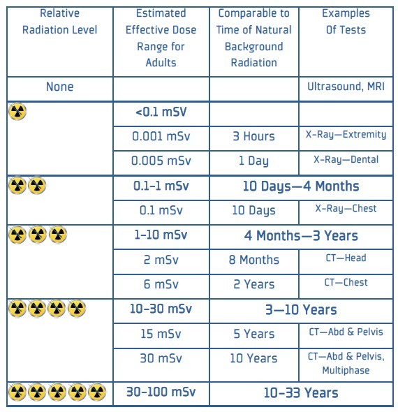
Medical imaging also causes our bodies to absorb radiation and the doses vary by test. See the next section regarding a variety of medical imaging tests and their relative radiation doses.
Medical Imaging and Radiation
The amount of radiation received from medical imaging tests varies according to the test, the testing protocols, length of the test and the testing equipment. However, estimates for the various tests will give you a better idea as to how much radiation exposure occurs with each test and in relationship to different tests.
Another way to look at the relative amount of radiation per imaging test is to compare tests to a reference amount of radiation, such as the amount of radiation from a chest X-ray.


